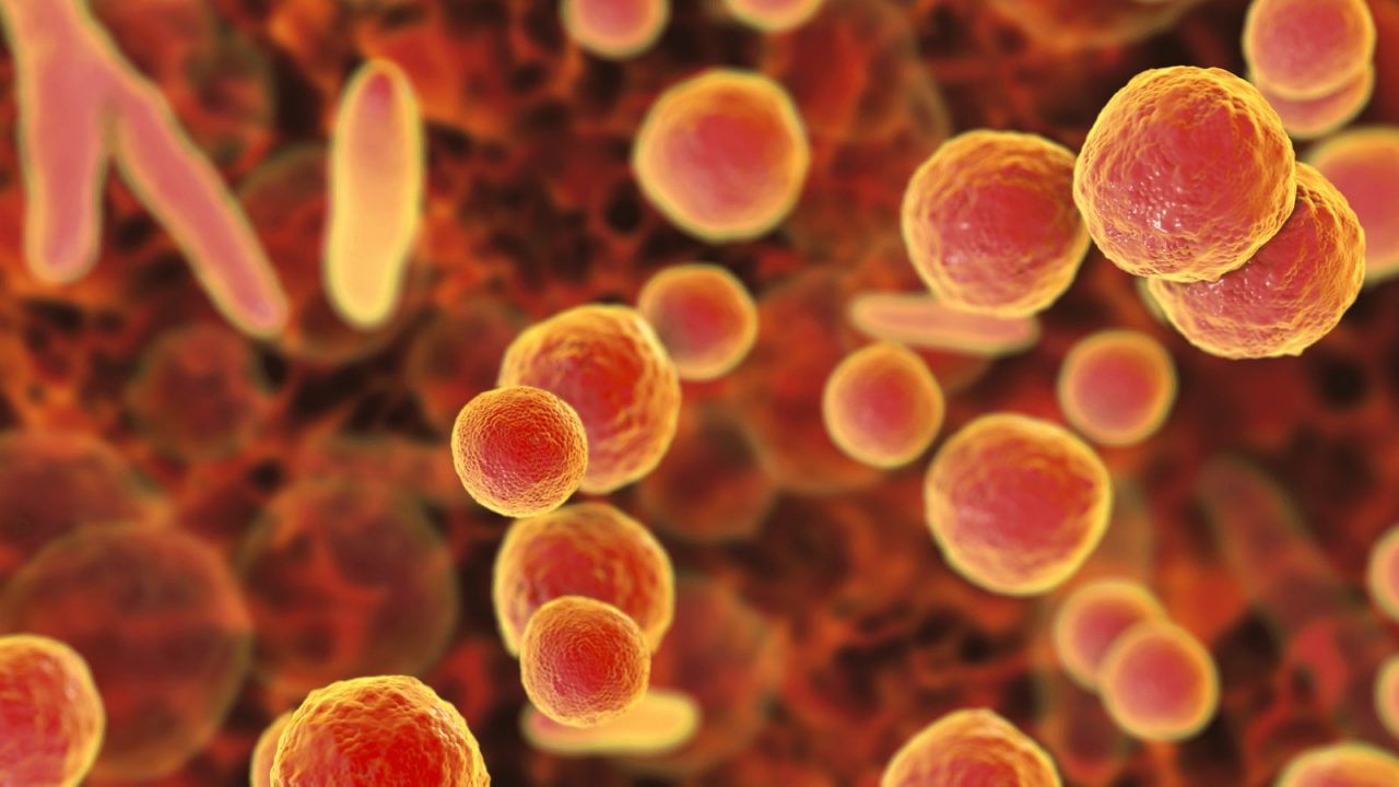
Mycoplasma hyorhinis
Potential Process Contaminant of Cell Culture Media

WHAT IS MYCOPLASMA HYORHINIS?
Mycoplasma is a genus of gram negative bacilli belonging to the Mollicutes class. Mollicutes include Mycoplasma and Acholeplasma. Mollicutes lack a cell wall and are of very small size (200-300 nm) and have a small genome. Since Mollicutes cannot produce many of the enzymes needed to survive they are parasitic on eukaryotic cells. Mycoplasma hyorhinis has mainly been associated with domestic pigs and rarely with human skin (where it has been associated with Scleroderma). It is associated with a couple of porcine diseases including enzootic pneumonia and porcine reproductive and respiratory syndrome (PRRS).
Given its widespread occurrence in nature it has been implicated as a common cell culture media contaminant of all cell culture media types. The top five cell culture contaminants (95%) have been identified as including: Mycoplasma orale, Mycoplasma arginini, Mycoplasma hyorinis, Mycoplasma fermentans, Acholeplasma laidlawii, or Mycoplasma hominis 1.
The small size and fluid structure of Mollicutes, in lacking a cell wall, contributes to their ability to evade the detection and control methods that are suitable for containing other microorganism types.
Mollicutes lack a cell wall and can penetrate sterilising filters under high pressure including 0.1 mm filters. The very low numbers which may pass through sterilising filters increase the probability that the contamination will not be detected during post production testing (Windsor, Windsor and Noordergraaf).
This article seeks to give an overview of Mycoplasma hyorhinis characteristics, many of which it shares with other Mycoplasmas, and suggests measures that can be used to ensure their proper detection and control.
HOW DOES MYCOPLASMA HYORHINIS COME TO BE A POTENTIAL PROCESS CONTAMINANT?
Mycoplasma hyorhinis can be present in raw materials of natural origin, particularly powder type additives to cell culture including bovine serum and trypticase soya broth. Mycoplasma hyorhinis growth in simple mycoplasma medium suggests it has minimal nutritional requirements. An increase in the use of filter sterilization for process production (in contrast to heat sterilization) has been viewed as a known association with the an increase in Acholeplasma laidlawii contamination events.
Mycoplasma and Acholeplasma contamination events can bring altered properties to cell cultures, resulting in the potential for altered cellular byproducts and even compromised biopharmaceuticals.
HOW CAN MYCOPLASMA HYORHINIS BE PREVENTED IN CELL CULTURE MEDIA?
The significant occurrence of mycoplasmas, including Mycoplasma hyorhinis, in cell cultures performed, a workhorse technology of the biotechnology field, can be seen in the estimates of 15 to 35% contamination rate of all cell culture processes 2.
Isolation of Mycoplasmas has less often been associated with the use of serum-free media 3, 4. Thus, where possible, serum-free medium can be chosen. Additional precautions include the use of heat sterilized, rather than filtered, water for reconstitution of powders included in media. Laidlaw and Elford found that cultures could not withstand 55 °C for 5 min and were significantly reduced in numbers after 15 min at 45 °C. Windsor, Windsor, and Noordergraaf confirmed these results where the survival of Acholeplasma laidlawii was reduced to zero in twenty minutes at 50 °C and 60 °C but not at 40 °C. According to these same researchers: “…it is not presently known how A. laidlawii becomes part of the bioburden of powders, or if similar powders (peptones or albumen) are the only potential source of this contaminant in serum free cell culture media.” And, “The ability of this organism to breach sterilising filters and then to grow to high titre in simple peptone broths or cell culture medium formulations stored at refrigeration temperatures has serious implications for the biopharmaceutical industry.”
HOW CAN THE PRESENCE OF MYCOPLASMA HYORHINIS BE DETECTED IN THE PHARMACEUTICAL INDUSTRY?
Testing for such contaminants in the pharmaceutical manufacturing environment is a universal requirement of regulatory agencies. Guidance for this testing is provided in the United States Pharmacopeia (USP) Chapter <63> Mycoplasma Tests, European Pharmacopoeia (EP) Chapter 2.6.7 Mycoplasmas, FDA 1993 Points to Consider (PTC), and the Code of Federal Regulations 21 CFR 610.30 test for mycoplasma.
Mycoplasma type contaminants such as Mycoplasma hyorhinis can go easily unnoticed as they remain invisible to the naked eye and light microscopy even at elevated titers. Routine testing is needed to ensure cell cultures are not contaminated and even that subsequent batches are not further infected from an originally contaminated batch. Detection methods include: direct growth on broth/agar, specific DNA staining, PCR, ELISA, RNA labeling and enzymatic procedures.
There are a number of technologies available on the market for detection of mycoplasmas in general and each contains associated advantages and disadvantages. The advantage category includes a broad coverage of Mollicute detection and a specificity for each species detected. Low false positive results are also important. Disadvantaged methods will provide false positives, will be time and labor consuming, and lack the desired specificity.
Given the sequencing of the complete genome of Mycoplasma hyorhinis 5 and other Mollicutes, PCR probes allow for their rapid and specific detection. The new BioFire® Mycoplasma panel for mycoplasma detection, includes a primer for M. hyorhinis as well as a full panel of other Mollicutes. Test sample preparation and test performance is simple and automated and results are ready in less than one hour.
HOW CAN MYCOPLASMA HYORHINIS BE FURTHER CONTROLLED IN THE BIOPHARMACEUTICAL INDUSTRY?
Chandler, Volokhov and Chizhikov claim that M. hyorhinis, and perhaps this concern should extend to all Mollicutes, may be prone to contaminate peptones and should be specifically precluded.
In general, vaccines and recombinant products produced in bacteria or yeast have not been tested for mycoplasmas, primarily because those production organisms grow rapidly in media that would not be expected to support the replication of the slow-growing mycoplasmas. There is one caveat, however, that is based on the recent reports that peptones (e.g., tryptone soya products sterilized by filtration) used in microbiological and cell culture media can be contaminated with viable mycoplasmas, specifically, M. hyorhinis 6. This observation makes it important to carefully evaluate all raw materials for potential mycoplasma contamination or to have procedures in place that will mitigate the risk of raw materials containing viable mycoplasma, such as pre-treatment of raw materials with ultraviolet radiation.
Industry control guidelines (or control measures) provide recommendations on how best to effectively control Mycoplasma hyorhinis e.g., in media, sera and process constituents. Emphasis is placed on selective purchasing with a documented supplier management program, Good Manufacturing Practices (GMPs), Hazard Analysis and Critical Control Points (HACCP), prerequisite programs, and robust continuous improvement activities.
These are means for all plants to use in accomplishing the goal of Mycoplasma hyorhinis reduction. Emphasis is on Raw Materials Purchasing Practices, Ingredient Shipping/Receiving, Physical Facilities, Plant Employees and Visitors, Plant Procedures and Policies (Sanitation and Cleaning, Pest Control, Materials Handling, Dust Control and Air Flow, Moisture Control), Equipment Maintenance and Operation, Packaging, Storage and Transportation, Control procedures, Sampling and Analysis.
WHAT COMMON INDUSTRIES ARE AFFECTED BY MYCOPLASMA HYORHINIS?
Mycoplasma hyorhinis is significant to all biopharmaceutical sectors employing cell culture techniques.
BIOMÉRIEUX SOLUTIONS AND PRODUCTS
BioFire® Mycoplasma panel
The BioFire® Mycoplasma Panel is a closed and fully-automated “lab in a pouch” system that provides highly sensitive and broad detection of Mollicutes with less than five minutes of hands-on time. The disposable reagent pouch integrates nucleic acid purification and nested multiplex real-time PCR with less than one hour of run time and requires minimal training and skill to operate.
Fifteen different PCR assays are included in every run to cover the wide genetic diversity of the Mollicutes class, including species of the genera Acholeplasma, Entomoplasma, Mesoplasma, Mycoplasma, Phytoplasma, Spiroplasma, and Ureaplasma. Three internal controls ensure the reliability of results by monitoring all aspects of pouch function, from sample extraction through amplicon detection.
Designed to meet compendial testing standards, Limit of Detection (LoD) studies have been performed with a particular focus on species listed by the United States, European, and Japanese pharmacopeias using 0.2 mL and 10 mL sample volumes. The LoD for 10 mL samples is ≤ 10 CFU/mL for all mycoplasmas tested and only requires a few simple pre-processing steps by centrifugation. Direct testing of 0.2 mL samples provides detection of ≤ 33 CFU/mL and is intended to facilitate more frequent surveillance of raw materials, cell banks, early product intermediates, and biologics with limited volume availability such as cell therapies.
REFERENCES
1 Considerations for risk and control of mycoplasma in bioprocessing, Angart et al., Current Opinionin Chemical Engineering, 2018,22:161–166.
2 The scope of mycoplasma contamination within the biopharmaceutical industry, Armstrong, Mariano , Lundin., Biologicals. 2010 Mar;38(2):211-3. doi: 10.1016/j.biologicals.2010.03.002.
3 Low IE. Isolation of Acholeplasma laidlawii from commercial serum free tissue culture medium and studies on its survival and detection. Appl Microbiol 1974:1046–52.
4 Beardsley T. Contamination at flow labs. Nature 1983;304:674.
5 Complete Genome and Proteome of Acholeplasma laidlawii, Lazarev et al., Journal of 5 Bacteriology Aug 2011, 193 (18) 4943-4953; DOI: 10.1128/JB.05059-11.
6 PDA (2010) Technical Report No. 50: Alternative Methods for Mycoplasma Testing. Bethesda, MD.


